


About breast biopsy
How is breast cancer diagnosed?
The process of diagnosing breast cancer includes three steps: an examination, imaging, and tissue sampling, where the final step is crucial.
- In the initial a breast examination, a doctor inspects both breasts and axillary lymph nodes and feels for any lumps or other abnormalities (so-called clinical examination).
- The second step employs mammography or other imaging diagnostics such as ultrasound, computed tomography, or MRI (magnetic resonance imaging).
- The third and crucial step for arriving at a correct diagnosis is the biopsy (tissue sampling).
Are mammography and ultrasound not sufficient for making a diagnosis?
No, the doctor cannot determine if a lesion is benign or cancer only by performing the physical examination. Mammography and ultrasound are good methods, but they alone do not provide a reliable answer. To confidently diagnose breast cancer, an analysis of cells and/or tissue from the suspicious area is needed. Hence, all lesions must be analysed based on cell or tissue samples.
What is a breast biopsy?
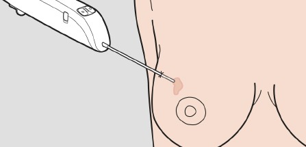
Biopsy means tissue sample. Breast biopsy is the removal of tissue or cells from a suspicious area in the breast.
Why are biopsies needed?
Breast biopsy is the only definitive diagnostic procedure that can determine if the suspicious area is cancerous. Apart from a reliable breast cancer diagnosis, biopsies also allow for:
- Determining the type of cancer.
- Determining the extent or size of a lesion.
- Optimising an individualised treatment regime.
- Monitoring the treatment and changing treatment type, if necessary.
How is a breast biopsy performed?
Previously, biopsies were performed surgically with an incision. Nowadays, breast biopsies are usually performed by a radiologist using a less invasive procedure, involving a hollow needle aided by a form of image guidance (for example ultrasound) to ensure that the sample is collected from the correct area.
How is a breast biopsy analysed?
The tissue sample is sent to a pathologist who analyses it (tissue structure and cells) under a microscope. The microscopic tissue analysis by the pathologist plays a central role in the diagnosis of cancer diseases.
Who performs needle biopsies of suspicious lesions?
In the UK, image-guided, minimally invasive procedures such as ultrasound-guided breast biopsy is are most often performed by a specially trained radiologist. This varies across different countries, of course.
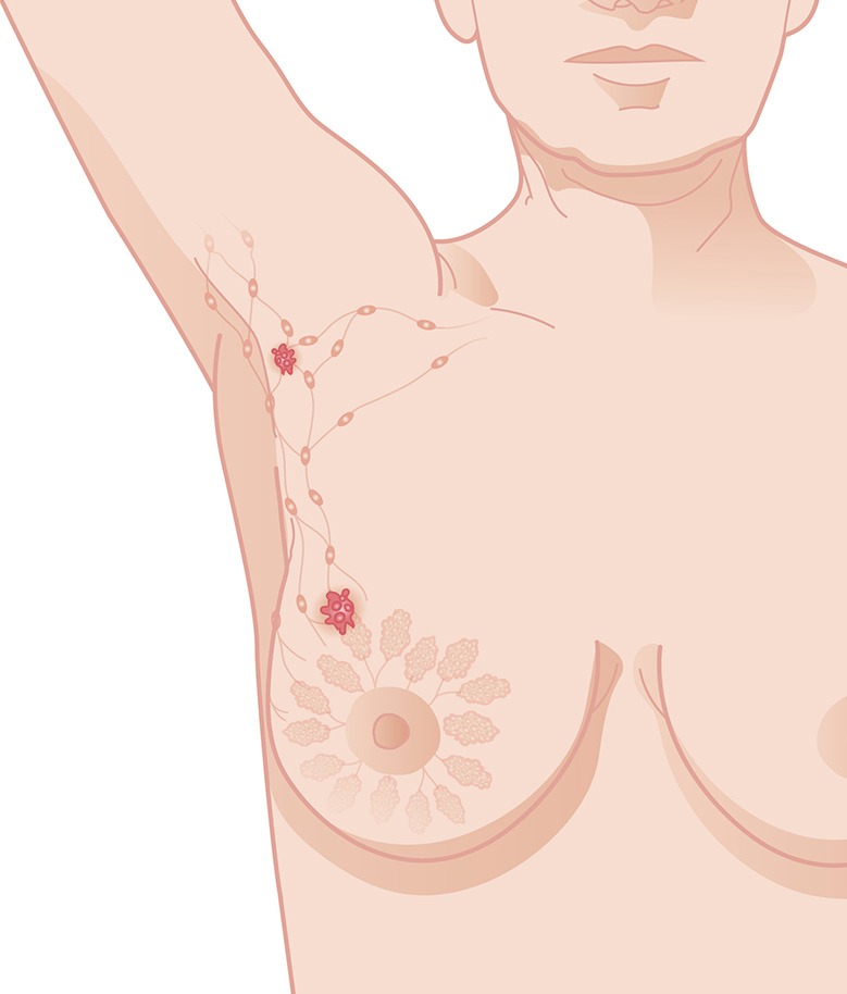
Why is axillary biopsy performed on breast cancer patients?
In the axillary lymph nodes, the lymph is drained from a tumour area. The spread of breast cancer usually first occurs to the lymph nodes in the armpit.
If one of the lymph nodes in the axilla appears suspicious on ultrasound, the doctor will take the opportunity and directly take a tissue or cell sample with a needle. An early examination of axillary lymph nodes provides accurate information regarding lymph node metastasis and thus the prognosis and allows for planning the treatment optimally even before any operation is performed. These samples are also taken by a radiologist or gynaecologist and analysed the same way as samples from the breast.
3 step diagnosis
- Physical examination
- Diagnostic imaging
- Tissue sampling
Breast biopsy is tissue sampling from the breast.
Sentinel Node biopsy can spare patients unnecessary discomfort after an operation.
